
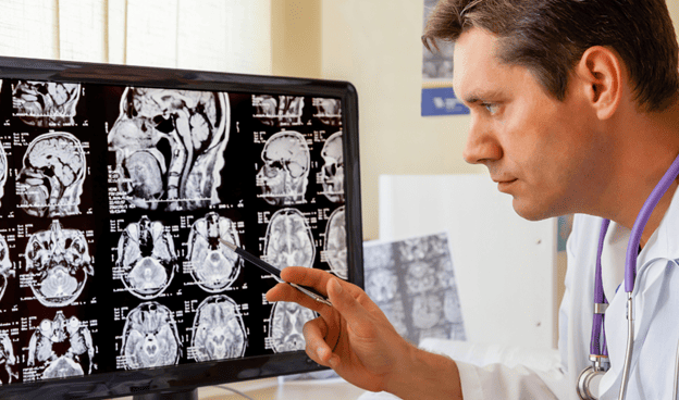
After a first seizure or unexplained episode, one of the biggest questions is often what caused it. A brain MRI can help answer part of that question by producing detailed images of the brain’s structure. Neurologists may use MRI to look for abnormalities that could contribute to seizures, including tumors, vascular abnormalities, scarring, developmental abnormalities, or evidence of previous brain injury.
MRI is only one part of a seizure evaluation. Some people with epilepsy have normal structural brain imaging, so a normal MRI does not rule out epilepsy. Neurologists may also consider what happened during the episode, medical history, neurological examination, EEG findings, and other diagnostic information.
For adults and families in River Forest and the greater Chicago area, CNS Brain Center provides neurological evaluation for seizures and other neurological concerns, with Neuro Imaging coming to its Chicago and River Forest locations.
Why Is a Brain MRI Used for Seizures?
A brain MRI allows neurologists to examine brain anatomy and look for structural abnormalities that could help explain or contribute to a seizure.
Depending on the patient’s history, neurologists may look for:
- Brain tumors or other masses
- Evidence of previous stroke or brain injury
- Vascular abnormalities
- Areas of scarring or tissue loss
- Developmental abnormalities
- Other lesions involving brain tissue
Finding an abnormality does not automatically mean it caused the seizure. Its location and characteristics must be considered alongside the patient’s symptoms, seizure history, EEG findings, and other clinical information.
Also Read: Brain MRI in Chicago: A Complete Guide to Advanced Neurological Imaging
What Does a Brain MRI Show After a Seizure?
What appears on an MRI after a seizure depends on the type of seizure, when the scan is performed, and whether an underlying structural abnormality is present.
Neurologists may evaluate brain anatomy and symmetry, areas of abnormal tissue, signs of previous injury, masses, vascular abnormalities, and structural changes in regions that may be relevant to the patient’s seizure pattern.
MRI can sometimes also show changes associated with a recent seizure. This means an abnormal finding may represent an underlying structural problem, a change related to the seizure itself, or something unrelated that requires further evaluation.
That is why MRI results need to be interpreted within the patient’s broader neurological picture rather than treated as a diagnosis on their own.
What Neurologists Look For on a Seizure MRI
Some abnormalities receive particular attention during seizure imaging because their location or characteristics may provide clues about why seizures are occurring.
Hippocampal and Temporal-Lobe Abnormalities
The hippocampus and temporal lobes are important in the evaluation of some forms of focal epilepsy. Neurologists may examine these areas for changes in structure or signal that could be relevant to the patient’s seizure pattern.
High-resolution epilepsy MRI protocols can provide detailed views of these regions, which can be important when abnormalities are subtle.
Developmental Changes, Scarring, and Previous Injury
Differences in cortical development can sometimes be associated with seizures. Neurologists may also look for areas of scarring or tissue loss related to previous trauma, infection, stroke, or another brain injury.
Whether one of these findings is significant depends partly on where it is located and whether it corresponds with other findings from the patient’s evaluation.
Tumors, Masses, and Vascular Abnormalities
MRI can identify tumors, other masses, and certain abnormalities involving blood vessels that may be relevant to seizures. If something unexpected is identified, additional imaging, testing, or specialist evaluation may be necessary to determine its significance.
Why MRI Detail Matters in a Seizure Evaluation
Some structural abnormalities associated with epilepsy can be difficult to see on routine imaging. For that reason, both image quality and the MRI protocol can matter when a scan is being performed specifically for seizures.
A 3T MRI uses a higher magnetic field strength than a 1.5T MRI. The additional signal available at 3T can support higher-resolution imaging and detailed visualization of brain anatomy. This can be particularly valuable when clinicians are evaluating subtle structural abnormalities.
The International League Against Epilepsy’s HARNESS-MRI recommendations for epilepsy imaging are optimized for 3T MRI when available. The protocol incorporates high-resolution sequences designed to examine brain anatomy and areas such as the hippocampus in greater detail. International League Against Epilepsy MRI recommendations
Not every patient who experiences a seizure needs the same MRI protocol. The appropriate imaging approach depends on the clinical circumstances and what the neurologist is trying to determine.
Also Read: EEG Testing: How It Helps Diagnose Neurological Conditions
Can a Brain MRI Detect Epilepsy?
MRI does not independently diagnose epilepsy. It primarily evaluates the structure of the brain, while an EEG records electrical brain activity.
These tests answer different questions:
- MRI: Is there a structural abnormality that could be relevant to the seizures?
- EEG: Is there abnormal electrical activity that may support an epilepsy diagnosis?
- Clinical history: What happened during the episode, and does it fit a seizure pattern?
A person can have epilepsy even when MRI does not reveal a structural abnormality. Likewise, an abnormal MRI finding does not automatically establish an epilepsy diagnosis.
Neurologists interpret these pieces of information together to determine what the findings may mean for the individual patient.
What Happens After a Seizure MRI?
What happens next depends on what the MRI shows and the rest of the neurological evaluation.
If a potentially relevant structural abnormality is identified, the neurologist may recommend additional imaging, testing, specialist evaluation, or treatment planning. If the MRI is normal, the evaluation may continue because some causes of seizures are not visible on structural imaging.
Depending on the situation, a seizure workup may include a medical and seizure history, neurological examination, EEG, laboratory testing, brain imaging, and other testing when clinically appropriate.
A normal MRI should therefore be interpreted as one piece of information, not as proof that a seizure did not occur.
Getting a Brain MRI for Seizures in River Forest
When arranging brain imaging for seizures, the MRI protocol and clinical indication matter. Patients may want to ask whether a seizure- or epilepsy-focused protocol is appropriate and how the results will be incorporated into their neurological evaluation.
Before MRI, patients should disclose implanted devices, previous surgeries, metal fragments, and other potential MRI safety concerns. Anyone who experiences claustrophobia should also discuss it with the imaging team before the examination.
CNS Brain Center’s Neuro Imaging service is being developed around advanced neurological imaging, including 3T MRI. CNS currently lists Neuro Imaging as coming to its River Forest and Chicago locations in Fall 2026. Patients can contact the center for current information about availability and scheduling.
Neurological Evaluation for Seizures at CNS Brain Center
Brain imaging is most useful when it is connected to the rest of the neurological evaluation rather than interpreted in isolation.
CNS Brain Center provides neurological evaluation and diagnostic testing for patients in River Forest, Chicago, and surrounding communities. For someone who has experienced a first seizure, recurrent episodes, or an unexplained event that could represent a seizure, a neurologist can determine whether EEG, MRI, or other testing is appropriate.
With advanced Neuro Imaging planned for its River Forest and Chicago locations, CNS is expanding its ability to evaluate neurological conditions within a comprehensive neurological setting.
Also Read: Understanding Your Neurological Health: EMG and NCV Tests in Chicago
Frequently Asked Questions
Why Is a Brain MRI Used for Seizures?
A brain MRI looks for structural abnormalities that may contribute to seizures, including tumors, vascular abnormalities, developmental changes, scarring, or evidence of previous brain injury. It is one component of a broader seizure evaluation.
What Is the Difference Between an MRI and an EEG for Seizures?
MRI provides detailed images of brain structure, while EEG records the brain’s electrical activity. Because they provide different information, neurologists may use both when evaluating suspected seizures or epilepsy.
Can an MRI Be Normal if Someone Has Epilepsy?
Yes. Some people with epilepsy have no structural abnormality visible on MRI. A normal scan therefore does not rule out epilepsy.
Can an MRI Show the Cause of a Seizure?
Sometimes. MRI may identify a structural abnormality that could contribute to seizures, but not every seizure has a cause that can be seen on structural imaging.
Why Might a 3T MRI Be Used for Epilepsy Imaging?
A 3T MRI can provide higher-resolution images and detailed visualization of brain anatomy. This may be useful when neurologists are looking for subtle structural abnormalities associated with epilepsy.
Where Can I Get a Brain MRI for Seizures in River Forest?
CNS Brain Center lists Neuro Imaging as coming to its River Forest and Chicago locations. Patients can contact the center for current information about 3T MRI availability and scheduling.
Schedule a Neurological Evaluation at CNS Brain Center
A brain MRI can provide valuable information after a seizure, but understanding what happened often requires looking at imaging alongside the patient’s history, neurological examination, EEG findings, and other appropriate testing.
If you have experienced a first seizure, recurrent seizures, or an unexplained episode, CNS Brain Center provides comprehensive neurological evaluation in River Forest and Chicago. Contact CNS Brain Center to schedule an evaluation or learn more about its Neuro Imaging services.