
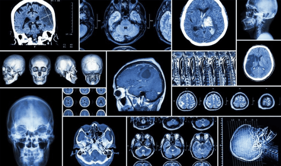
New or worsening memory problems can raise difficult questions for patients and their families. You may wonder whether the changes are related to normal aging, a neurological condition, medication, vascular disease, or another medical problem. A brain MRI can provide detailed images of brain structure and help physicians look for abnormalities that may contribute to cognitive symptoms.
Understanding what MRI can and cannot reveal is an important part of a memory-loss evaluation. MRI can show structural changes such as brain atrophy, evidence of prior stroke, certain vascular abnormalities, tumors, hydrocephalus, and other findings that may help guide the diagnostic process. It cannot, by itself, determine every cause of memory loss or establish every form of dementia.
For patients experiencing memory problems or cognitive decline, a neurological evaluation can combine the patient’s symptoms and medical history with cognitive testing, neurological examination, laboratory testing, and appropriate brain imaging.
Why Would a Doctor Order a Brain MRI for Memory Loss?
Memory changes can have many possible causes, and imaging is not necessary in every situation. A physician may recommend a brain MRI when symptoms are new, worsening, unusual, or accompanied by other neurological changes.
MRI may help physicians identify:
- Evidence of prior stroke or other vascular brain injury
- Changes associated with vascular disease
- Brain tumors
- Abnormal fluid accumulation, including hydrocephalus
- Areas of brain volume loss
- Other structural abnormalities
Brain scans are commonly used as one part of a broader dementia or memory evaluation. They can help identify potentially treatable causes and provide information about structural changes in the brain.
The decision to order imaging depends on the individual patient. A physician may consider the patient’s symptom history, physical and neurological examination, cognitive testing, medical conditions, medications, and other factors.
For this reason, it is too simplistic to say that an MRI for memory loss simply “looks for dementia.” MRI provides structural information that physicians interpret alongside symptoms, examination findings, and other testing to investigate possible causes of cognitive change.
Sudden confusion or memory change accompanied by weakness, numbness, facial drooping, speech difficulty, severe headache, loss of consciousness, or another abrupt neurological change requires urgent medical evaluation rather than a routine outpatient memory assessment.
Also Read: The Science of Brain Longevity: Tips for a Healthy Mind for Life
What Can a Brain MRI Reveal About Memory Loss?
MRI provides detailed images of brain anatomy and can reveal structural changes that may be relevant to cognitive symptoms.
One important finding is brain atrophy, or loss of brain volume. Some forms of dementia are associated with characteristic patterns of shrinkage in particular regions of the brain. The location and pattern of atrophy can provide useful diagnostic clues when interpreted alongside symptoms and other testing.
MRI can also show evidence of vascular injury. For example, imaging may reveal previous strokes or changes involving blood vessels and white matter that can contribute to cognitive impairment.
Other structural abnormalities may also be visible. MRI can help physicians look for tumors, fluid buildup, and other conditions that can cause symptoms resembling dementia or contribute to cognitive problems.
This means brain imaging for memory loss can be valuable even when the ultimate diagnosis is not dementia. An imaging study may help identify another explanation for a patient’s symptoms or provide information that changes the direction of the evaluation.
Can an MRI Detect Dementia or Alzheimer’s Disease?
MRI can reveal structural changes associated with some types of dementia and provide information that helps physicians investigate possible causes of cognitive decline. However, an MRI alone generally does not establish a dementia diagnosis.
- A physician may combine MRI findings with:
- Medical and family history
- Cognitive or neuropsychological testing
- Neurological examination
- Laboratory testing
- Medication review
- Other brain imaging or biomarker testing when appropriate
The National Institute on Aging describes dementia diagnosis as a process that can include medical history, physical and neurological testing, laboratory testing, cognitive assessment, and brain imaging.
So, can MRI show Alzheimer’s disease? MRI can show patterns of brain shrinkage that may support a clinical assessment of Alzheimer’s disease, but it is not generally a standalone test that confirms Alzheimer’s disease. There is no single simple test that establishes an Alzheimer’s diagnosis in every patient.
A normal MRI also does not necessarily rule out early cognitive disease. Structural changes may be subtle or not yet apparent, particularly during earlier stages.
What Brain Changes Can MRI Detect in Patients With Memory Problems?
MRI may reveal several types of structural changes that can be relevant to a memory evaluation:
Brain Volume Loss
Different neurological disorders can produce different patterns of brain volume loss. In Alzheimer’s disease, for example, changes involving the hippocampus and surrounding brain tissue may be visible as the disease progresses.
The presence of atrophy does not automatically establish a diagnosis. Physicians consider the location, degree, and pattern of volume loss together with the patient’s symptoms and other clinical findings.
Vascular Changes
MRI can identify evidence of strokes and other changes associated with damaged blood vessels. These findings can help physicians evaluate whether vascular disease may be contributing to cognitive symptoms.
White-Matter Abnormalities
Changes in the brain’s white matter can be associated with vascular disease and other neurological conditions. Their significance depends on the patient’s age, symptoms, medical history, and the pattern and extent of the abnormalities.
Other Structural Abnormalities
MRI can also help identify tumors, fluid buildup, and other structural conditions that can produce cognitive or neurological symptoms.
The important point is that an imaging finding should not be interpreted in isolation. Two people can have similar MRI findings but very different symptoms and diagnoses.
A normal or nonspecific MRI does not mean that a patient’s memory concerns are not real, nor does it completely exclude early cognitive disease. Structural imaging findings must be interpreted within the broader clinical evaluation.
Also Read: Medical-Grade HBOT for Neurodegenerative Conditions
What Does an MRI Show in Early Dementia?
Another common question is what does an MRI show in early dementia?
In some patients, MRI may show subtle patterns of brain volume loss or other structural changes that provide diagnostic clues. However, early dementia may not produce obvious changes on a conventional structural MRI, and a normal or nonspecific scan does not completely exclude early disease.
MRI is therefore best understood as one component of the evaluation rather than a standalone dementia test.
In some circumstances, physicians may consider additional testing when the diagnosis remains unclear. Depending on the clinical situation, this can include specialized brain imaging, laboratory studies, neuropsychological testing, or other biomarker assessments.
What Happens During a Brain MRI for Memory Loss?
If your physician recommends a brain MRI for memory loss, the process generally begins with an MRI safety screening.
The MRI team may ask about:
- Metal implants
- Pacemakers or other implanted devices
- Previous surgeries
- Metal fragments or foreign bodies
- Other conditions that may affect MRI safety
During the scan, you typically lie on a movable table while your head is positioned for imaging. The table moves into the MRI scanner, which uses magnetic fields and radio waves to create detailed images.
MRI machines can be loud, so patients may be provided with hearing protection. Remaining as still as possible during the scan is important because movement can reduce image quality.
Whether contrast material is needed depends on the clinical question and the MRI protocol selected by the ordering physician. Contrast is not automatically required for every evaluation of memory loss.
Your physician determines whether imaging is appropriate and what type of MRI protocol is needed based on your symptoms and clinical circumstances.
What Happens After the MRI?
After imaging is completed, a radiologist interprets the MRI images and prepares a report. The physician managing the patient’s care then considers those findings alongside the patient’s symptoms, medical history, neurological examination, and other test results.
An MRI finding can sometimes lead to additional testing or follow-up. In other cases, the scan may provide useful information by making certain structural explanations less likely.
It is also possible for an MRI to reveal an incidental finding that is unrelated to the patient’s memory symptoms. This is another reason patients should not attempt to interpret individual images without medical context.
A physician’s interpretation considers not only whether something appears abnormal but also whether the finding is clinically relevant to the patient’s symptoms.
Why Choose CNS Brain Center for Memory and Neurological Evaluation?
CNS Brain Center provides neurological care for patients experiencing memory changes and other neurological symptoms in the River Forest and Chicago areas.
When memory changes develop, a comprehensive neurological evaluation can help place the symptoms in context. Rather than relying on a single imaging finding, physicians can consider the patient’s history, symptoms, examination findings, cognitive concerns, and appropriate diagnostic testing.
The value of MRI in a dementia or memory evaluation comes from interpreting the images within the patient’s broader clinical picture rather than relying on the scan alone. CNS Brain Center can help determine what type of neurological evaluation or consultation may be appropriate based on the patient’s symptoms and medical history.
Neurological evaluation and MRI imaging may also occur at different locations. Patients should confirm imaging availability and scheduling with the appropriate medical facility.
Also Read: Your New Treatment Options for Alzheimer’s Dementia
Conclusion
A brain MRI can provide valuable information when a patient develops new or worsening memory problems. It may reveal brain atrophy, vascular changes, evidence of stroke, tumors, hydrocephalus, and other structural abnormalities that can help physicians investigate the cause of cognitive symptoms.
However, MRI cannot independently diagnose every form of dementia or explain every memory complaint. MRI for memory loss is most useful when interpreted alongside the patient’s symptoms, medical history, neurological examination, cognitive testing, and other appropriate diagnostic information.
If memory problems are persistent, worsening, or affecting daily life, a professional neurological evaluation can help determine what may be contributing to the changes and whether brain imaging or other testing is appropriate.
For patients in the River Forest and Chicago areas, CNS Brain Center provides individualized neurological evaluation and consultation.
Frequently Asked Questions
Can a brain MRI detect the cause of memory loss?
An MRI can help identify structural or vascular abnormalities that may contribute to memory loss, including evidence of prior stroke, tumors, hydrocephalus, and patterns of brain atrophy. However, MRI cannot identify every possible cause of memory loss by itself.
Can an MRI show Alzheimer’s disease?
MRI can show patterns of brain atrophy that may be consistent with Alzheimer’s disease and can identify structural or vascular findings that suggest other explanations for cognitive decline. However, MRI alone does not establish an Alzheimer’s diagnosis.
What happens during a brain MRI for memory problems?
During a brain MRI for memory problems, the patient typically lies on a movable table with the head positioned for imaging. The scanner can be loud, and remaining still is important for obtaining clear images. MRI safety screening is performed before the scan.
Where can I get a brain MRI or neurological evaluation near River Forest?
Patients in the River Forest area can contact CNS Brain Center to discuss neurological evaluation and consultation options. Whether MRI imaging is appropriate and where it should be performed depends on the patient’s medical circumstances and the physician’s evaluation.